
I published “Symptoms of Rectal Prolapse” on @Medium https://ift.tt/3kEGaIl


Based in New York, Dr. Brian Gilchrist is a longtime pediatric surgeon who has leadership experience in everything from trauma care to neonatal conditions. During his perinatology and obstetrics rounds at the State University of New York’s Health Science Center at Brooklyn, Dr. Brian Gilchrist spoke on “Management of Choledochal Cysts.”
Congenital, or present at birth, choledochal cysts can either appear in infancy or take several years to emerge. Among the symptoms to be aware of are abdominal mass, nausea, fever, and vomiting, as well as a pain in the upper right belly.
Since the liver generates bile as a digestive aid, choledochal cysts involve anomalies of the duct responsible for transporting bile from liver to small intestine and gall bladder. As the duct swells abnormally, bile backs up into the liver, which can, in turn, cause liver issues. It can also block the major duct leading from the pancreas gland to intestine, which may lead to pancreatitis, or inflammation of the pancreas.
The long-term risks of this occurring in childhood are significant, including an elevated cancer risk as an adult. The positive news is that choledochal cysts are a treatable condition and can be successfully removed through laparoscopic surgery.

Dr. Brian Gilchrist, a skilled pediatric surgeon with over 30 years of experience, has held many teaching roles and has been invited to give numerous presentations and lectures before medical professionals. Dedicated to advancing knowledge in the medical field, Dr. Brian Gilchrist edited a book about necrotizing enterocolitis (NEC).
A gastrointestinal disease that affects both premature and full-term infants, NEC is responsible for inflammation and infection in the intestines, leading to the damage and death of intestinal cells. The condition is categorized into three stages:
– Stage one is suspected NEC with mild symptoms such as lethargy and bloody stool;
– Stage two is definite NEC with the symptoms of stage one along with pain when the abdomen is touched and low blood platelet levels;
– Stage three is advanced NEC accompanied by low blood pressure and other issues.
Treatment of NEC depends on the stage, age, overall health, and tolerance to the medication. Surgery is usually required for stage three NEC. By this point, a hole may have developed in the bowel or intestine. Surgery removes the affected area and leaves undamaged intestines to regain their normal function.


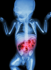
A member of the American Pediatric Surgical Association, Dr. Brian Gilchrist is a New York-based pediatric surgeon who has delivered lectures worldwide and has written and edited numerous publications on neonatal and pediatric surgical topics. Dr. Brian Gilchrist’s works include a book on necrotizing enterocolitis that is well-referenced in neonatal intensive care units across the country. Necrotizing enterocolitis (NEC) is a life-threatening intestinal disease with a high mortality rate in newborns and is the most frequent gastrointestinal emergency in NICUs.
The condition, which is characterized by inflammation and the bacterial invasion of the large intestine, most commonly affects severely ill and premature infants, particularly ones who weigh less than 3 pounds, 4 ounces. Intestinal tissue damage and death can occur as result of the inflammation and bacteria, which can create a hole in the intestines and destroy its walls. Symptoms usually appear within two weeks after a baby’s birth and can include bloody bowel movements, stomach bloating, fever, green bile, and poor feeding tolerance. Lethargy, slow heart rate, and pauses in breathing can also indicate infection.
Despite continuous research, the specific cause of NEC remains unknown. Medical professionals believe that low blood and oxygen flow play a part because the disease is most prevalent in premature infants who are born with weaker and immature lungs and intestines. These babies also have trouble fighting infection and breaking down food. In addition to premature birth, factors such as intestinal infections, difficult birth, and formula feeding for high-risk and premature infants can heighten the risk of NEC.
Healthcare providers can diagnose NEC by examining babies for signs of the condition and conducting an abdominal X-ray. A bubbly appearance in the intestine and indicators of gas or air in a child’s large veins in the liver are NEC indicators that can show up in an X-ray. Treatment can vary due to multiple factors, such as the severity of the disease and the child’s symptoms, general health, and age. Common steps can include halting feedings, providing intravenous fluids and nutrition, and offering oxygen support and using breathing machines.
